

Researchers have developed an AI tool that reveals previously unseen patterns inside breast cancers.
A University of Southampton team, working with the city’s hospital, used the technology to analyse more than 330,000 cells from 127 patients.
The findings could help doctors identify high-risk patients and develop more targeted treatments.
Leading the research, the university’s Dr Salah Elias, said: “This opens the door to developing new biomarkers and, ultimately, more personalised treatment strategies.”
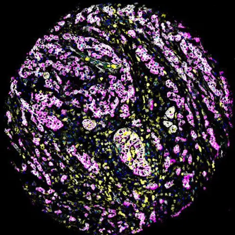
The study found two different types of changes inside cancer cells that had previously been treated as the same thing.
One type involved cells developing too many centrosomes, which help cells divide properly. The other involved centrosomes becoming unusually large.
Researchers discovered the two changes could appear in different parts of the same tumour and may play different roles in how cancers develop.
The team created an AI platform, called CenSegNet, to examine tumour samples with “unprecedented speed and precision”.
Centrosomes act as the cell’s organising centres, helping cells divide and maintain their structure. When they become abnormal, cells can build up genetic mistakes, which are commonly seen in cancer.
The researchers found tumours with higher numbers of enlarged centrosomes were more likely to have features linked to more aggressive disease.
These included higher tumour grade, cancer that had spread to nearby lymph nodes and certain genetic changes.
The study also found patients with fewer enlarged centrosomes in the centre of their tumours tended to have better overall survival.
Elias, said: “For more than a century, centrosome abnormalities have been recognised as a hallmark of cancer, but studying them in patient tissues has been extremely challenging.
“CenSegNet allows us to analyse these defects at single-cell resolution across entire tumours and uncover patterns that were previously impossible to see.”
Researchers say the technology could eventually help doctors better understand which cancers are more likely to grow, spread or resist treatment.
It could also help scientists develop more personalised treatments by identifying tumours with specific weaknesses that could be targeted with new drugs.
While the technology is not yet ready for routine use in hospitals, the team has also shown it can be used on tissue samples from other parts of the body, including the kidney, colon and appendix.
CenSegNet is available free of charge as open-source software, allowing researchers around the world to use and study the technology.

